

Date: June 12, 2025
Dr. Alons Lends
Affiliation: Latvian Institute of Organic Synthesis
Talk Title: “Breaking the Resolution and Sensitivity Limits of Intact Fungal Cell Walls with Proton-Detected Solid-State NMR at 60–150 kHz MAS”
Dr. Lends, a leading expert in the field of magnetic resonance, presented his latest research on overcoming the inherent challenges of studying complex and heterogeneous fungal cell walls. His talk emphasized the power of high-frequency, proton-detected solid-state NMR (SSNMR) to achieve unprecedented levels of resolution and sensitivity, even in living samples.
Key Takeaways and Innovations:
- High-Resolution Insights into Complex Cell Walls
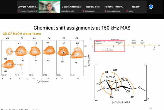
By employing proton-detected SSNMR at spinning rates of up to 150 kHz, Dr. Lends and his team have significantly improved the spectral resolution achievable in heterogeneous fungal samples. This advancement allows for the detailed detection and analysis of crucial structural components, such as 1,3-β-glucans, which are essential for the integrity and function of fungal cell walls. Such high-resolution insights were previously unattainable, marking a major leap forward in the field.
- Studying Living Cells Inside the NMR Rotor
A remarkable aspect of this research is the demonstration that deuterated fungal cells can remain viable for more than two weeks while packed inside the MAS rotors. This finding opens exciting new possibilities for conducting non-invasive, real-time studies of living microbial systems, enabling researchers to observe biological processes as they happen, without disrupting cell viability or function.
- Investigating Drug Interactions in Real Time
The use of fluorine-labeled antifungal compounds as sensitive NMR probes has enabled the direct observation of drug-cell wall interactions. Changes in chemical shifts provided clear evidence of drug binding to specific cell wall components. Additionally, Dr. Lends discussed the potential for further signal enhancement using Dynamic Nuclear Polarization (DNP), which could make these experiments even more powerful and informative in the future.
- Structural Characterization with Precision
By applying J-coupling measurements, the research team successfully distinguished between α- and β-anomers of polysaccharides within the cell wall. This capability allows for reliable and detailed spectral assignments of native cell wall components, facilitating a deeper understanding of fungal cell wall architecture at the atomic level.
- Practical Considerations for Researchers
The seminar also addressed practical aspects of the methodology. Samples were packed in 0.51 mm MAS rotors using specialized tools from Ago, and it was noted that these rotors are reusable, with only the end caps needing replacement—a cost-effective solution with replacement costs estimated at €1000–1500. The workflow is fully compatible with ultracentrifugation protocols, making it accessible and convenient for laboratories worldwide.
During the interactive discussion, participants explored the differences between fixed and non-fixed samples, noting that only protonated samples were fixed while deuterated samples remained metabolically active. The team confirmed the viability of cells under extended MAS spinning conditions, and practical questions regarding rotor recycling, cost, and handling were thoroughly addressed.
Significance and Impact
This presentation clearly demonstrated how advanced SSNMR techniques are now enabling researchers to:
- Conduct structural studies of living fungal cells at atomic resolution, providing insights that were previously out of reach.
- Monitor drug-cell wall interactions in real time, which is invaluable for drug discovery and development.
- Expand the application of solid-state NMR into new areas of microbiology and pharmaceutical research, bridging gaps between structural biology and applied sciences.
Thank you to all attendees and especially Dr. Alons Lends for a compelling presentation!